

Advanced Eye Care Technology
We choose technology for one reason: better care. Our diagnostic tools help us capture more detailed measurements, make the exam more comfortable, and give your doctor more time to focus on you.
Better Technology Means a Better Exam Experience
Advanced imaging and diagnostic tools allow us to see more than a standard exam can reveal. We can detect concerns earlier, follow subtle changes over time, and better understand your long-term eye health, while keeping the visit comfortable and efficient.
Digital Wide-Field Retinal Imaging
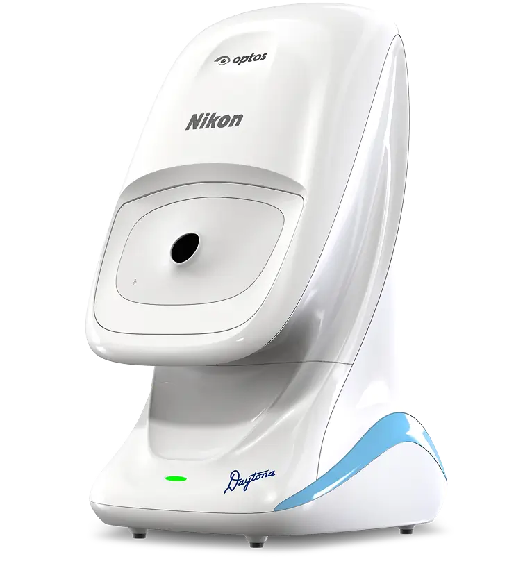
Optomap
Optomap captures a wide, high-resolution image of the retina in seconds, giving our doctors a more complete view of the back of the eye.
This helps us detect and monitor subtle changes over time, including signs of macular degeneration, diabetic eye disease, optic nerve changes, and suspicious retinal freckles or lesions.
Many eye conditions develop quietly. Detailed imaging helps us see more before symptoms appear.

Ocular Neuro-Alignment
Newton
Newton uses advanced virtual reality testing to measure how your eyes align and work together.
Subtle misalignments can contribute to headaches, eye strain, motion sensitivity, neck tension, light sensitivity, or dry eye symptoms. By identifying these issues, Newton helps guide non-invasive treatment options for clearer, more comfortable vision.
Eye Pressure
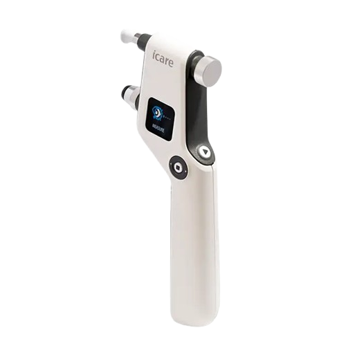
iCare Tonometer
The iCare Tonometer measures eye pressure quickly and gently, without the sudden burst of air used in traditional puff tests.
This helps make glaucoma screening more comfortable while giving our doctors reliable information to monitor eye health over time.
Corneal and Tear Film Imaging
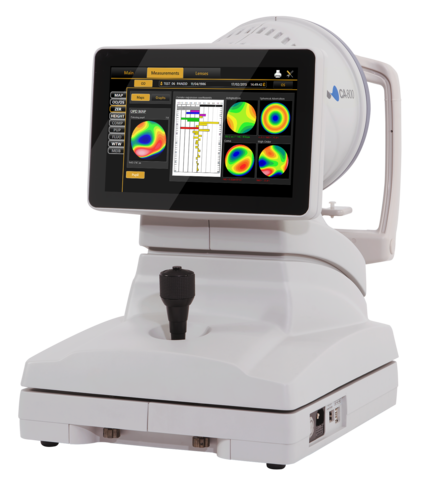
Topography and Meibography
Topography maps the shape of the cornea, while meibography shows the oil glands that help keep your tears stable and comfortable.
Together, these tools help us better understand dry eye, meibomian gland dysfunction, keratoconus, and other corneal conditions, so treatment can be more targeted and changes can be monitored over time.

Macula and Optic Nerve Imaging
Optical Coherence Tomography (OCT)
OCT gives us a detailed, layered view of the macula and optic nerve, revealing structures that cannot be seen through a standard exam alone.
With this level of imaging, our doctors can better detect, diagnose, and monitor conditions such as glaucoma, macular degeneration, retinal changes, and other concerns that may affect long-term vision.
Our Standard
- Cutting-edge, modern diagnostic technology
- Full-view, HD retinal mapping
- Biometric eye alignment
- Gentle, non-invasive pressure testing
The Standard
- Legacy hardware
- Narrow, low resolution imaging
- Manual alignment estimates
- Standard “air-puff”
Ready to Book Your Exam?
Schedule your visit and experience technology chosen to make your care more detailed, comfortable, and personal.